

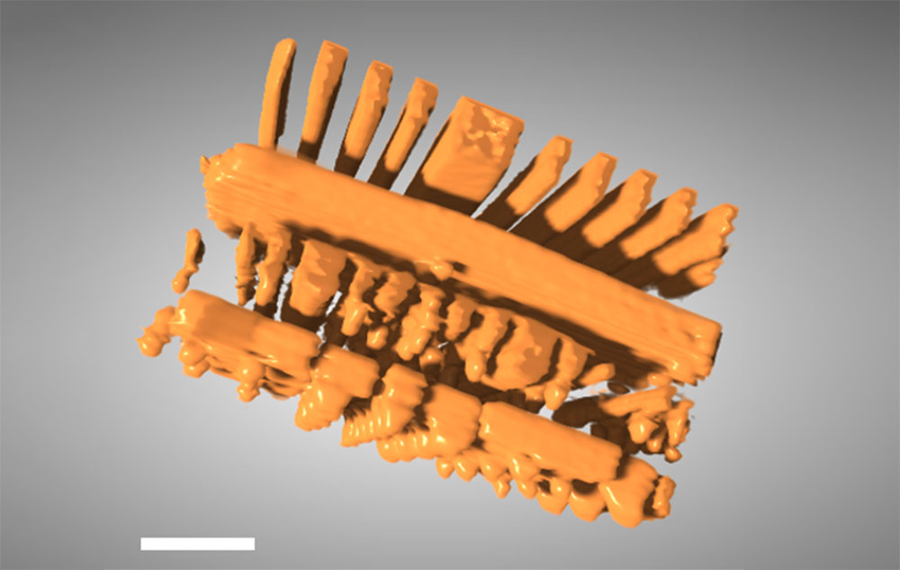
Scientists at the Hard X-ray Nanoprobe beamline at the National Synchrotron Light Source II (NSLS-II), a U.S. Department of Energy user facility at Brookhaven National Laboratory, have created a new artificial intelligence-assisted approach that significantly improves three-dimensional X-ray tomography. Traditional X-ray tomography works by rotating a sample and collecting projection images from all angles, but in real applications such as imaging flat or densely packed objects, some angles are inaccessible. The missing information from those angles creates a “blind spot” known as the missing wedge, which leads to blurry and distorted reconstructions. The new method directly addresses that problem.
The technique, dubbed the perception fused iterative tomography reconstruction engine (PFITRE), tightly couples a convolutional neural network with a physics-based model of X-ray imaging. The AI component, trained on synthetic and simulated data, captures perceptual insights about typical image features such as edges, curves, and textures, while the physics model ensures that results remain faithful to the underlying measurements. By iterating between these two elements, PFITRE yields reconstructions that are both visually sharp and scientifically accurate, even when a full set of angular projections cannot be collected.
The Brookhaven team designed PFITRE so the neural network works as a “smart regularizer” within an iterative solver, improving detail without sacrificing physical consistency. The AI is based on a U-net architecture enhanced with structural innovations that improve how features are detected and synthesized. Because real experimental datasets are limited, the researchers trained the model with virtual data that mimics real conditions, including noise and misalignment.
The advance expands what’s possible with nanoscale imaging. PFITRE makes it feasible to extract detailed internal views of samples that were previously difficult or impossible to image because of geometry or data limitations. Beyond sharper images, the method could enable faster experiments, require fewer measurements, and reduce radiation exposure for sensitive materials. Its authors see applications in fields ranging from microelectronics to battery research and materials science.
