

Medical ultrasound is one of the most widely used imaging techniques in healthcare, but interpreting its images can be challenging. Clinicians typically view a series of two-dimensional slices and must mentally reconstruct them into a three-dimensional understanding of a patient’s anatomy. This process requires significant training and experience, making ultrasound interpretation difficult for many users.
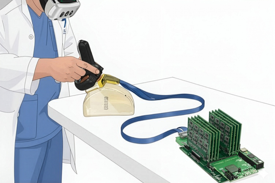
Researchers at the Massachusetts Institute of Technology have developed a new augmented reality (AR) ultrasound system designed to address this challenge. The technology allows users to see a real-time, three-dimensional digital representation of the object being scanned while performing an ultrasound examination. By wearing a virtual reality headset, clinicians can visualize anatomical structures as accurate 3D models rather than relying solely on conventional 2D images.
The system combines ultrasound data with advanced spatial tracking and visualization techniques to create a precise digital rendering of tissues and organs. Instead of mentally piecing together multiple image slices, users can observe the scanned structure directly in three dimensions, making it easier to identify features and understand spatial relationships. This approach has the potential to improve both diagnostic accuracy and workflow efficiency.
The researchers designed the system to project ultrasound information into the user’s field of view while maintaining alignment with the physical object being scanned. This creates a more intuitive imaging experience and enables users to interact with ultrasound data in a way that closely matches real-world anatomy. The technology could be particularly valuable in situations where rapid interpretation of imaging data is critical.
Beyond clinical practice, the system could significantly enhance ultrasound education and training. Because ultrasound imaging is highly operator-dependent, learning to interpret scans can take considerable time. The AR-based visualization approach may shorten that learning curve by helping trainees connect ultrasound signals with the anatomical structures they represent.
The researchers also see potential applications in image-guided procedures such as biopsies and other minimally invasive interventions that require precise instrument placement. By providing clearer spatial awareness, the technology could improve procedural accuracy and safety.
Overall, the project demonstrates how augmented reality can transform medical imaging by making ultrasound data more intuitive, accessible, and easier to interpret. The work highlights the growing role of immersive visualization technologies in improving healthcare delivery, medical training, and patient outcomes.
