Ants are among the most abundant and widespread organisms on Earth, with scientists estimating their population at roughly 20 quadrillion. Despite their tiny size, these insects play major ecological roles and exhibit remarkably complex social systems. A new scientific effort has now brought ants into unprecedented focus through a large collection of detailed three-dimensional images that reveal their anatomy both inside and out, tells The New York Times.
In a study published in Nature Methods, biodiversity scientist Evan Economo of the University of Maryland and his collaborators produced high-resolution 3D scans of nearly 2,200 ant specimens. The insects came from museums and private collections worldwide and were scanned during an intensive week at a synchrotron particle accelerator in Karlsruhe, Germany. This powerful facility enabled researchers to complete in days what would normally require years using conventional micro-CT scanning equipment.
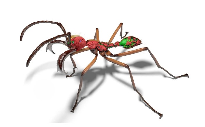
A synchrotron produces extremely bright X-ray beams, allowing scientists to image specimens rapidly while also visualizing soft tissues without chemical staining. As a result, the scans capture extraordinary detail, including internal organs such as brains, digestive systems, and glands. The resulting images showcase a remarkable diversity of forms, from large species such as the bullet ant to extremely small predators specialized in feeding on spider eggs.
Many preserved ants initially appeared contorted because specimens are typically stored in curled positions. To address this, computer scientists collaborating on the project used artificial intelligence to reposition the scans into natural poses. The technique helps create lifelike digital models that can be used for scientific analysis, education, and even artistic or animation projects.
The scans have already yielded scientific insights. For example, researchers confirmed that a mineralized armor previously identified in certain fungus-farming ants appears in many related species as well. Experts say the ability to examine organisms internally without physically dissecting them could transform biological research.
The digital models are freely available online at antscan.info, providing scientists and the public alike with access to an extraordinary visual archive. The project also points toward a broader ambition: building detailed digital representations of Earth’s biodiversity.